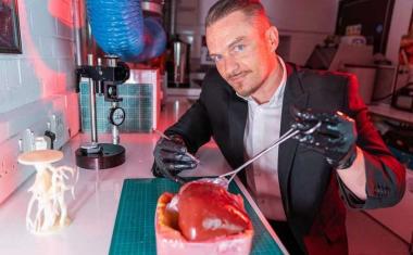
器官生物制动器呼吸新鲜空气
工程师们对巴勒斯坦权力机构清除了一个重要障碍th to 3D printing replacement organs with a breakthrough technique for bioprinting tissues. The new innovation allows scientists to create exquisitely entangled vascular networks that mimic the body’s natural passageways for blood, air, lymph and other vital fluids.
The work was led by bioengineers Jordan Miller of Rice University and Kelly Stevens of the University of Washington (UW) and included 15 collaborators from Rice, UW, Duke University, Rowan University and Nervous System, a design firm in Somerville, Massachusetts.
“One of the biggest road blocks to generating functional tissue replacements has been our inability to print the complex vasculature that can supply nutrients to densely populated tissues,” said Miller, assistant professor of bioengineering at Rice’s George R. Brown School of Engineering. “Further, our organs actually contain independent vascular networks – like the airways and blood vessels of the lung or the bile ducts and blood vessels in the liver. These interpenetrating networks are physically and biochemically entangled, and the architecture itself is intimately related to tissue function. Ours is the first bioprinting technology that addresses the challenge of multivascularization in a direct and comprehensive way.”
广告

广告


Stevens, assistant professor of bioengineering in the UW College of Engineering, assistant professor of pathology in the UW School of Medicine, and an investigator at the UW Medicine Institute for Stem Cell and Regenerative Medicine, said multivascularization is important because form and function often go hand in hand. “Tissue engineering has struggled with this for a generation,” Stevens said. “With this work we can now better ask, ‘If we can print tissues that look and now even breathe more like the healthy tissues in our bodies, will they also then functionally behave more like those tissues?’ This is an important question, because how well a bioprinted tissue functions will affect how successful it will be as a therapy.”
生物印刷的目标健康,功能器官是由于器官移植的需要驱动。超过10万人在美国独自移植等候名单,最终接受捐赠器官的人仍然面临免疫抑制药物的一生,以防止器官排斥。Bioplinting在过去十年中引起了激烈的兴趣,因为它可以理论上通过允许医生从患者自己的细胞打印替换器官来解决这两个问题。随时可以部署有一天的功能器官,以治疗全球数百万患者。“我们希望在未来二十年内成为医药的主要成分,”米勒说。
“The liver is especially interesting because it performs a mind-boggling 500 functions, likely second only to the brain,” Stevens said. “The liver’s complexity means there is currently no machine or therapy that can replace all its functions when it fails. Bioprinted human organs might someday supply that therapy.”

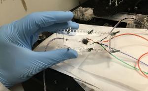
To address this challenge, the team created a new open-source bioprinting technology dubbed the “stereolithography apparatus for tissue engineering,” or SLATE. The system uses additive manufacturing to make soft hydrogels one layer at a time.
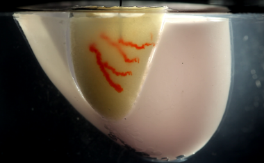
从液体前水凝胶溶液中印刷层,当暴露于蓝光时,将成为固体。数字光处理投影机从下面闪耀光,在高分辨率下显示序列2D结构,像素尺寸范围为10-50微米。
当依次凝固的每个层依次凝固,架空臂升高了生长的3D凝胶,足以将液体暴露于从投影仪的下一个图像。Miller和Bagrat Grigoryan的主要洞察力,米饭研究生和该研究的领导共同作者,加入吸收蓝光的食物染料。这些光吸收器将凝固物限制在非常细的层中。通过这种方式,该系统可以在几分钟内产生具有复杂内部架构的软,水基的生物相容性凝胶。
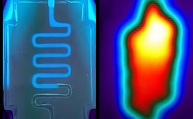
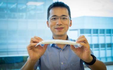
Tests of the lung-mimicking structure showed that the tissues were sturdy enough to avoid bursting during blood flow and pulsatile “breathing,” a rhythmic intake and outflow of air that simulated the pressures and frequencies of human breathing. Tests found that red blood cells could take up oxygen as they flowed through a network of blood vessels surrounding the “breathing” air sac. This movement of oxygen is similar to the gas exchange that occurs in the lung’s alveolar air sacs.
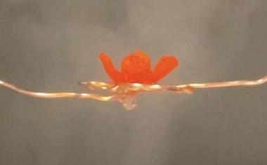
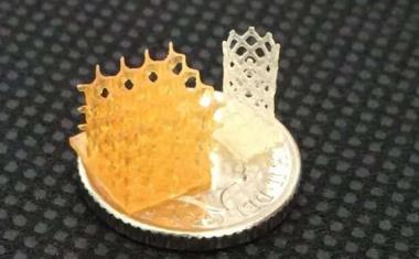
设计该研究最复杂的肺模拟结构,该结构在科学封面上,米勒与学习共同作者Jessica Rosenkrantz和Jesse Louis-Rosenberg,神经系统的联合创始人。“当我们创立紧张的系统时,它是目的,目的是将算法从大自然中调整为新的设计产品,”Rosenkrantz说。“我们从未想过我们有机会带来背部和设计生活组织。”
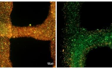
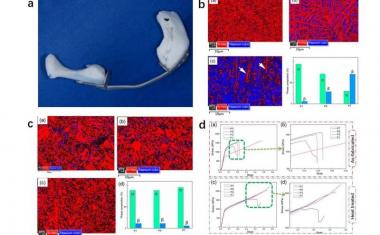
在肝病治疗植入物的试验中,三维3D印刷组织,用原发性肝细胞加载它们并将它们植入小鼠。组织有血管和肝细胞的单独隔室,并植入慢性肝损伤的小鼠中。试验表明肝细胞存活植入。
米勒说,新的生物打印系统还可以公关oduce intravascular features, like bicuspid valves that allow fluid to flow in only one direction. In humans, intravascular valves are found in the heart, leg veins and complementary networks like the lymphatic system that have no pump to drive flow. “With the addition of multivascular and intravascular structure, we’re introducing an extensive set of design freedoms for engineering living tissue,” Miller said. “We now have the freedom to build many of the intricate structures found in the body.”
Miller和Grigoryan正在通过呼佣的初创公司公司商业化研究的关键方面,称为体积。Grigoryan加入全职的公司,正在设计和制造生生物制剂和生物链条。
Miller, a longstanding champion of open-source 3D printing, said all source data from the experiments in the published Science study are freely available. In addition, all 3D printable files needed to build the stereolithography printing apparatus are available, as are the design files for printing each of the hydrogels used in the study. “Making the hydrogel design files available will allow others to explore our efforts here, even if they utilize some future 3D printing technology that doesn’t exist today,” Miller said.
Miller said his lab is already using the new design and bioprinting techniques to explore even more complex structures. “We are only at the beginning of our exploration of the architectures found in the human body,” he said. “We still have so much more to learn.”
Source:Rice University