Researchers successfully bioprint healthy new tissue
New muscle has successfully been created in mice using a minimally invasive technique dubbed ‘intravital 3D bioprinting’ by a team involving University College London scientists.
This new research could pave the way for minimally invasivesurgicaltechniques for organ repair and reconstruction that could remove the need for transplantation in children with complex conditions.
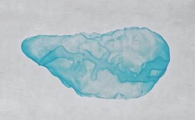
For pioneering international study, researchers from the UCL Great Ormond Street Institute of Child Health developed a photosensitive bio-gel that uses light treatment to ‘print’ healthy newtissuedirectly into specific tissues and organs, and sustain blood supply that would allow it to thrive.
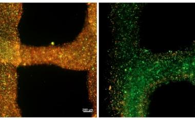
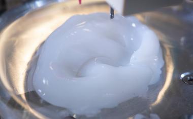
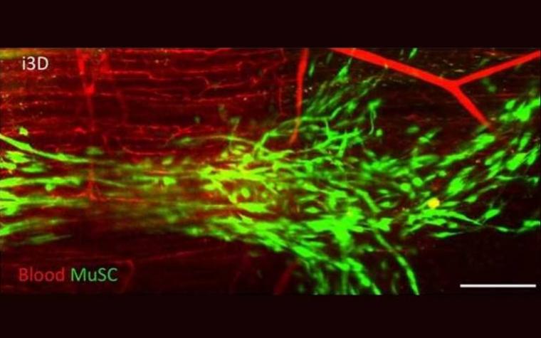
The light-sensitive bio-gel acted as a type of bio-ink, effectively ‘printing’ 3D structures that supported the creation of muscle fibres in the muscle of live mice and without the need for open surgery.
Advertisement

Advertisement

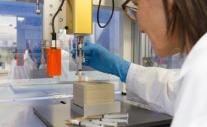
Researchers loaded a liquid gel with cells carefully selected to suit the type of tissue being printed. The bio-gel was then injected into the area of interest in the body with a simple syringe. Once in place, the team directed a near infrared light at the area from outside the body. Polymers inside the bio-gel bonded together under this wavelength of light, solidifying 3D structures layer by layer and allowing the cells to reach the desired position. With support from the structures, the cells adapted and connected to their new surroundings to form new tissue.
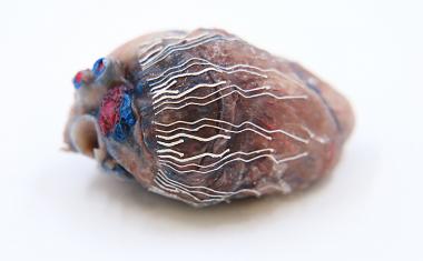
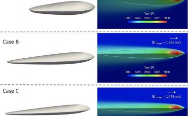
Initially tested on the skin and brain of the mouse, the team’s technique – coined Intravital新利18官方, or i3D Bioprinting – was also successfully carried out in the muscle of a mouse where it created new tissue without causing damage to surrounding organs or tissues.
Furthermore, it did not create any waste material inside the body and has the potential to carry healthy donor cells. This could be life-changing in cases where a child’s own cells aren’t suitable or available to help repair or reconstruct damaged or missing tissue.
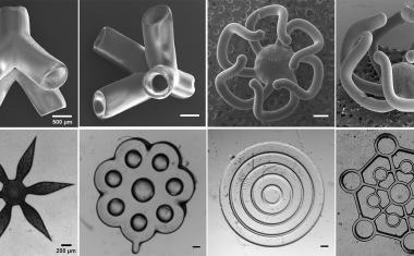
Project lead Professor Nicola Elvassore (UCL GOS Institute of Child Health), whose research team spans the ICH, Italy and China, said: “Recent attempts at 3D bioprinting have required direct access to the tissue and space to manoeuvre the 3Dbioprinting笔,如何控制它的形状和组织形式structure. That meant they focused on parts of the body that are easier to access, such as the skin. We’re really excited that our technique seems to be much more controllable in three dimensions, allowing us to accurately image in 3D the anatomical sites of interest and safely print new tissue in areas that aren’t easy to access without major surgery, like the brain.”
Recommended article
3D printed biomaterial enables forming of blood vessels
An international team of scientists have discovered a new material that can be 3D printed to create tissue-like vascular structures. In a new study, researchers have developed a way to 3D print graphene oxide with a protein which can organise into tubular structures that replicate some properties of vascular tissue.
First-author Dr Anna Urciuolo, visiting research associate at the UCL GOS Institute of Child Health, said: “It was an exciting and challenging project, which required the fusion of emerging technologies in a multidisciplinary approach. By performing 3D bioprinting directly within the body of live animal model, we were able to deliver donor musclestem cellsin a spatially controlled manner, increasing their ability to develop new muscle tissue.”
The international team, which included researchers from Italy’s University of Padova and Veneto Institute of Molecular Medicine, successfully ‘printed’ the gel within skin, muscle and brain tissues. Co-author Professor Paolo De Coppi, who is Nuffield Professor of Surgery at UCL GOS Institute of Child Health and a Consultant at Great Ormond Street Hospital (GOSH), said: “This is an important step in repairing damaged tissue and offers the possibility of minimally invasive regeneration, which could change in the future the way we treat congenital malformations such as spina bifida and diaphragmatic hernia at GOSH. We still have plenty of work to do before we can safely use this approach with patients, but the pre-clinical findings are promising.”
Although still in its pre-clinical stage, this kind of tissue engineering research could lead to a new standard of care for patients with complex physical conditions, especially in the case of children with damagedorgans.
Source:University College London