How to make the invisible visible
Scientists have discovered a new way to analyse microscopic cells, tissues and other transparent specimens, through the improvement of an almost 100-year-old imaging technique.
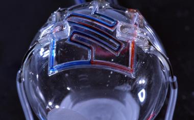
拉筹伯University researchers have led a four-year collaboration to make "the invisible visible" by using custom-designed nanomaterials to enhance the sensitivity ofphase contrast microscopy, an imaging technique commonly used by scientists to study biological specimens.
The discovery will benefit a broad range of researchers and has the potential to advance research into the understanding and detection of disease.
Advertisement

Advertisement

项目负责人和拉筹伯Molecula研究所r Science (LIMS) physicist, Professor Brian Abbey, said the discovery allows scientists to detect minute changes in the composition or structure of transparent or nano-thin objects, enabling their key features and structures to be visible when put under a microscope.
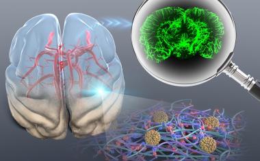
"Features that were previously impossible to detect using conventional techniques can now be imaged using our microscopy method, developed by lead author and La Trobe Senior Research Fellow Dr. Eugeniu Balaur," Professor Abbey said. "It has allowed our teams to delve deeper into the identification of early destructive processes inneurodegenerative diseases, such asmultiple sclerosis."
Phase contrast microscopy, an optical technique that can be utilized to produce high-contrast images of transparent specimens, was first invented in 1934 by Nobel-prize winning physicist Frits Zernike. "This technique allows scientists to examine cells in their natural state without previously being stained or labeled. As a result, their structure and function—and perhaps even their dynamics—can be better understood," Professor Abbey said. "We now have the tools to manipulate matter at the nanoscale. Our custom designed nanomaterials have enabled us to achieve a huge leap forward in terms of image quality and contrast, building on the groundbreaking phase work of Zernike in the 1930s."
拉筹伯neuroscientist and study co-author Dr. Jacqueline Orian said the research team is working to translate the study to ensure their new microscopy technique can be utilized by scientists worldwide. "A major application of the technique would be in the evaluation of candidate drugs for promotion of repair to nerves and their protective casings known as myelin," Dr. Orian said.
"The technique the team developed enables us to perform label-free imaging of cellular structures with exceptional contrast and in their natural state. Coupling nanotechnology with phase contrast microscopy lays the foundation for entirely new fields of research and represents a significant leap forward for the life sciences," Dr. Nicholas Anthony, study co-author from the Istituto Italiano di Tecnologia (IIT) said.
The research was published in自然光子学.
Source:拉筹伯University