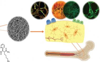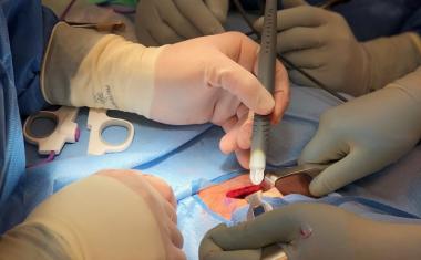
用纳米洛碳研究心脏细胞
一种新的微电极,渗透细胞膜,并且当放置在阵列中时,允许科学家遵循电活动,因为它通过组织传播。
Cells are the smallest living units in the human body. Excitable cells such as neurons and cardiac muscle cells – cardiomyocytes – use electrical signals, so-called action potentials, to communicate with each other. Scientists study these signals underlying normal brain and heart function using electrodes placed either outside or inside the cell membrane, methods known as extracellular and intracellular recording.
Researchers at EPFL’s Microsystems Laboratory 4 (LMIS4), led by Philippe Renaud, and the Laboratory of Cellular Optics II of the University of Bern, headed by Stephan Rohr, have teamed up to develop a new microelectrode that penetrates the cell membrane unassisted and, when placed in an array, allows scientists to follow electrical activity as it spreads through tissues.
多年来,虽然蜂窝电气活动的记录系统显着发展,但它们仍然有局限性。非侵入性细胞外α阵列,其使用放置在膜报告信号外部的电极仅与动作电位间接相关的信号。他们告诉科学家对动作电位的实际形状几乎没有 - 例如细胞的瞬时升高 - 例如,这导致心脏击败。
Since cellular action potentials were first measured by Silvio Weidmann from the University of Bern’s Department of Physiology seven decades ago, scientists have been measuring these signals by gaining intracellular access with microelectrodes. These electrodes can be impaled into the cells, or they can be placed onto the cell membrane, after which the membrane is opened under the mouth of the electrode. This can be done either mechanically or by electroporation – the application of high voltage pulses to the electrode. The latter technique was recently used to gain intracellular access by nanostructured electrodes in the shape of microscopic mushrooms, for example. However, this methodology is not ideal because the interface between the cell membrane and the nanostructure is unstable, leaving only a brief window – typically a few seconds or minutes at most – for scientists to record action potentials from cells.
广告

广告

受到自然的启发
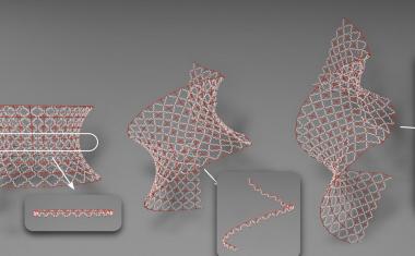
The EPFL and University of Bern team took the best features of existing technologies and came up with an ingenious volcano-shaped design to get around this problem. “By reworking the geometry and materials, we developed an electrode that penetrates the cell membrane unassisted, thus eliminating the need for electroporation,” says Benoît Desbiolles, a doctoral assistant at LMIS4 and lead author of the publication. “We also drew on previous research by our lab, which shows that mimicking the cell membrane stabilizes the cell-electrode interface.”
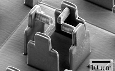
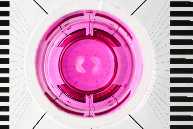
The new type of electrode, coined as a nanovolcano, consists of three parts. The first is the rim of the crater. It consists of a gold ring having the same size and being lined with the same biomolecules as the cell membrane itself. Inside the crater sits a platinum electrode used for picking up the electrical signals. The outside is surrounded by insulating glass. “Once you place a cell on the structure and it starts to bed down, the sharp edges pierce the membrane and the electrode penetrates the cell,” explains Desbiolles. “Instead of reforming, the membrane anchors itself to the gold ring, creating the ideal conditions to record the cell’s electrical activity.”
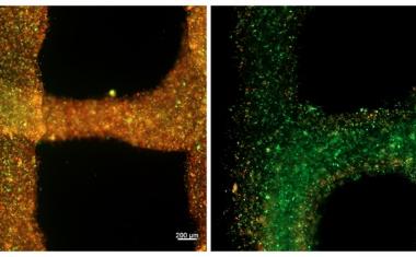
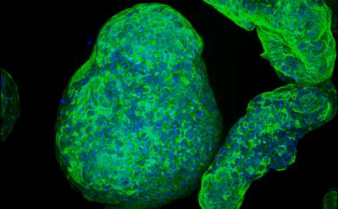
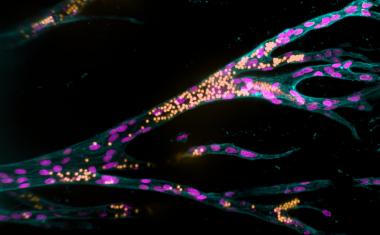
使用Nanovolcano阵列,科学家可以同时测量细胞培养中的多个位置的动作电位,为心肌细胞如何在空间中相互作用,提供了丰富的见解。“对于像我这样的电生理学家来说,这项技术是一个梦想成真的东西,”斯蒂芬·罗尔说,他共同撰写了该出版物。“以及测量单个细胞的动作潜力,我们现在可以研究传播动作电位如何根据组织结构和病理条件改变它们的形状。这种知识对于更深入地了解导致可能致命的心律失常的机制至关重要。“
纳米洛西州的潜在应用远远超出心脏电生理学。“除了其开创性设计之外,我们的电极也非常容易制作,”Desbiolles解释道。目前正在运行测试,看看它是否与神经元和其他易激的细胞类型同样好。根据年轻的研究人员,该设计持有对其他科学学科的承诺:“纳沃尔卡诺在电池中打开门。您可以在内部可想地执行电化学。“该技术也可能吸引制药行业,允许科学家测试细胞如何对毒品的反应,从而长期地发展有针对性的疗法。